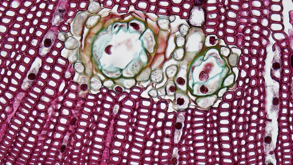
Introduction
Histology is crucial in medicine and science. It helps us understand tissue structure and function. Tissue sampling plays a vital role in diagnosing diseases. By examining tissues under a microscope, we can identify abnormalities. Understanding histological principles enhances effective tissue analysis and improves patient outcomes.
Summary and Overview
Tissue sampling histology involves collecting, preserving, and analyzing tissue samples. The primary objectives include ensuring preservation of tissue integrity, preparing samples for examination, and analyzing them to detect diseases. Proper techniques are essential for accurate diagnoses. These techniques can significantly impact treatment options, guiding physicians in their decisions. By utilizing histological methods, we gain insights into various conditions, leading to better patient care.
For those interested in further exploration, numerous PDF resources are available. These documents provide valuable information and can deepen your understanding of histological principles. They are excellent references for students, researchers, and professionals in the field. Utilizing these resources can enhance your knowledge and improve your skills in tissue sampling and analysis.
If you’re looking for a solid foundation in histology, consider picking up Essential Histology. This textbook is designed for medical students and provides a thorough overview of histological techniques and concepts, ensuring you won’t miss a beat in your studies!
For a comprehensive understanding, you can refer to the add embedding data to seurat guide.
Understanding Histology
What is Histology?
Histology is the study of microscopic structures in tissues and organs. It plays a significant role in medical diagnosis and research. By examining tissues at a cellular level, we can uncover details that inform us about health and disease. Histology bridges the gap between structure and function, providing insights into how tissues work and what happens when they malfunction. This understanding is vital for developing effective treatments and therapies in various medical fields.
If you’re diving deeper into histology, you might want to check out Histology: A Textbook for Medical Students. This textbook provides in-depth knowledge about histological techniques and is perfect for both students and professionals looking to refresh their skills.

Tissue Preparation Process
Fixation
Fixation is the first step in tissue preparation. It preserves the tissue’s natural structure and prevents decay. This process stabilizes proteins and halts enzymatic reactions. Without fixation, tissues would degrade quickly, leading to inaccurate results. Common fixatives include formaldehyde and glutaraldehyde. These chemicals cross-link proteins, forming a gel-like matrix that maintains cellular integrity. Proper fixation is crucial for successful staining and microscopic analysis. It ensures that the tissue remains representative of its in vivo state.
To achieve optimal fixation, you might consider using a Histological Stains: A Practical Manual. This manual gives you the practical tips you need for choosing and using the right stains, making your histology work shine!

Dehydration
After fixation, the next step is dehydration. This process removes water from the tissue, which is essential for embedding. Ethanol is typically used for this purpose, as it effectively replaces water in the tissue. The dehydration process usually occurs in graded ethanol solutions, starting from low concentrations to 100% ethanol. This gradual increase prevents tissue shrinkage and damage. After dehydration, xylene may be used to remove ethanol, preparing the tissue for embedding in paraffin or resin.
Embedding
Embedding is a critical phase in tissue preparation. In this step, the dehydrated tissue is infiltrated with a solid medium, usually paraffin wax. This creates a stable matrix that allows for precise sectioning. Proper embedding ensures that the tissue is oriented correctly, which is vital for obtaining quality sections. When the paraffin hardens, it forms a block that can be easily sliced into thin sections. These sections are crucial for histological examination. Without proper embedding, the tissue may not cut well, leading to poor-quality slides.
For those looking to set up a histology lab, consider investing in a Histology Lab Equipment Set. This comprehensive set will cover all your basic needs, ensuring you’re well-equipped for your histological adventures!
Diagnostic Staining Techniques
Common Stains Used in Histology
Hematoxylin and Eosin (H&E)
Hematoxylin and Eosin (H&E) is the gold standard in histological staining. This process involves two key steps. First, hematoxylin stains nuclei a deep blue or purple, highlighting DNA and RNA-rich areas. Next, eosin imparts a pink hue to the cytoplasm, emphasizing protein-rich structures. Together, these stains provide a clear contrast between cellular components, making it easier to identify abnormalities. H&E staining is crucial for diagnosing various conditions, including cancer, as it reveals the architecture of tissues.
For those interested in enhancing their staining skills, you might want to grab a Hematoxylin and Eosin Stain Kit. This kit makes it easier to perform H&E staining with confidence, ensuring your slides look professional!

Gram Stain
The Gram staining technique is essential in microbiology for classifying bacteria. It consists of four steps. First, the specimen is stained with crystal violet, which colors all cells. Next, iodine is added to form a complex with the crystal violet. The third step involves decolorization, usually with alcohol, which washes out the dye from Gram-negative bacteria. Finally, a counterstain, safranin, is applied, turning Gram-negative bacteria pink. This differentiation helps determine the appropriate antibiotics for treatment, influencing patient care significantly.
Special Stains
Special stains target specific components within tissues and provide detailed insights. For example, the Periodic Acid Schiff (PAS) stain highlights carbohydrates. It turns structures like glycogen and mucins a vibrant red or magenta. This is particularly useful in diagnosing conditions such as diabetes or fungal infections.
Masson’s Trichrome stain is another specialized technique that differentiates between collagen and muscle fibers, giving collagen a blue color and muscle a red hue. This stain is particularly helpful in examining fibrotic tissues and understanding scarring.
Other notable special stains include:
- Giemsa Stain: Useful in hematology for identifying blood parasites.
- Congo Red: Stains amyloid deposits, appearing red with green birefringence under polarized light, aiding in diagnosing amyloidosis.
- Silver Stain: Highlights nerve fibers and certain proteins, crucial in neurological studies.
These specialized stains play a pivotal role in histopathology by providing information that standard stains might miss. They enhance our understanding of tissue pathology, leading to better clinical outcomes. Each stain serves a unique purpose, allowing pathologists to tailor their approach based on the suspected condition.
To make your staining process even easier, check out the Gram Stain Kit. This kit provides all the necessary reagents to perform the Gram staining process seamlessly!

Microscopy in Histology
Types of Microscopes
Light Microscopy
Light microscopy is a fundamental tool in histology. It uses visible light to illuminate specimens, allowing for the observation of living or fixed tissues. When light passes through the sample, it magnifies the image, making it possible to see structures within cells. This technique typically achieves magnifications up to 1500 times.
Light microscopy is ideal for routine histological examinations. It highlights cellular details and allows pathologists to identify abnormalities. Stains like Hematoxylin and Eosin (H&E) enhance contrast, revealing intricate structures in tissue sections. This method is widely used in laboratories for diagnosing diseases, as it provides clear imaging of tissue architecture.
If you’re looking to upgrade your microscopy skills, consider getting a Light Microscope. It’s an essential piece of equipment for anyone serious about histology, offering excellent clarity and precision for your observations!

Electron Microscopy
Electron microscopy offers a powerful alternative for examining tissues at much higher magnifications. Instead of light, it uses a beam of electrons to create images. This allows for magnifications up to 200,000 times, revealing fine details of cellular structures.
The main advantage of electron microscopy lies in its ability to visualize ultrastructural components, such as organelles and membranes. This is crucial in understanding pathological changes at a cellular level. For instance, it can reveal the presence of viral particles or other abnormalities that light microscopy may miss. Therefore, electron microscopy plays a pivotal role in research and diagnostics, particularly in complex cases requiring detailed analysis.
For those ready to take the plunge into advanced microscopy, a Electron Microscope might be your next best investment. This tool allows you to explore the microscopic world in stunning detail!
Image Analysis
Image analysis tools have revolutionized histological evaluation. Software applications assist pathologists in quantifying and analyzing tissue samples. They enable precise measurements of cell size, density, and morphology. This quantitative approach enhances objectivity and accuracy in diagnoses.
Digital pathology is a growing field that integrates imaging technology with pathology. It allows for the storage and sharing of high-resolution images. Pathologists can review slides remotely, improving collaboration and consultation. Moreover, artificial intelligence is increasingly being used to assist in image analysis, helping to identify patterns and anomalies that may not be immediately apparent. This modern approach significantly enhances the efficiency and effectiveness of histological methods.

Clinical Applications of Histology
Importance in Disease Diagnosis
Histology plays a vital role in diagnosing various diseases. By examining tissue samples, pathologists can identify conditions such as cancer, infections, and autoimmune disorders. For example, in cancer diagnosis, histological techniques reveal cellular abnormalities that indicate malignancy. The identification of tumor type and grade is essential for determining treatment options.
Implications for Treatment
The findings from histological analyses significantly influence treatment decisions. For instance, the type of cancer identified through histology can determine the course of therapy, such as surgery, chemotherapy, or targeted treatments. Histology also informs prognosis, helping physicians predict disease progression and patient outcomes.
As personalized medicine becomes more prevalent, histological analysis is becoming even more critical. It allows for tailored treatment plans based on individual patient profiles and tumor characteristics. This personalized approach to medicine enhances the effectiveness of treatments and improves overall patient care.
For those interested in enhancing their histology knowledge, consider getting a Histology Reference Guide. This guide can be a handy resource for quick consultations and clarifications during your studies!

FAQs
What is tissue sampling histology?
Tissue sampling histology involves collecting, preserving, and analyzing tissue samples to diagnose diseases. It provides critical insights into tissue structure and function.
Why is fixation important in histology?
Fixation is vital as it preserves tissue structure. It prevents decay and deformation, ensuring accurate analysis during microscopic examination.
What are the common staining techniques used in histology?
Common staining techniques include Hematoxylin and Eosin (H&E), Gram stain, and special stains like the Periodic Acid Schiff (PAS) stain. Each technique highlights different cellular components for diagnosis.
How does microscopy enhance tissue analysis?
Microscopy enhances tissue analysis by allowing detailed observation of tissue structures. Light and electron microscopy reveal cell morphology and ultrastructure, aiding in accurate diagnoses.
What role does histology play in disease diagnosis?
Histology is essential in diagnosing diseases by identifying cellular abnormalities. It provides crucial information for determining conditions like cancer, infections, and autoimmune disorders.
Please let us know what you think about our content by leaving a comment down below!
Thank you for reading till here 🙂
All images from Pexels


